AI for medical reporting
Better reports
in less time
Create reports for radiology, nuclear medicine, cardiology and other specialties from your dictation. Laudos.AI follows your template. You review and sign.
14 days or 30 reports free · no card
52smedianRadiology dataFrom editor to signature04/09–05/09/2026 · in productionFrom exam to report
You interpret the examThe report, we write together
Every word in this lung comes from the report. Hover to read it. You read the image; Laudos.AI writes the report from what you dictate.
Results in radiology
52 secondsfrom editor to signature
of reports in under 1 minute
from opening the editor to signature.
recovered per 100 reports
of radiologist time.
fidelity to the signed report
across 120 cases of snapshot v3.10.
versioned classifications
served by API. No score goes out without a source.
Median measured in production from April 9 to May 9, 2026; it does not measure total TAT or clinical quality. LaiBench is an internal benchmark, not external clinical validation.
Made by people who report
Less repetitive workMore attention to the diagnosis
You assess the exam, decide and dictate. Laudos.AI organizes the findings in your report template, with no commands or memorized phrases.
See the demoIndication, history and priors arrive from the RIS and the PACS, with no manual upload.
You assess the exam and dictate. Technique, findings and conclusion follow your specialty's format.
The report goes back to the RIS/PACS, with an auditable trail and the critical finding recorded.
“
Low-dose computed tomography of the chest, without intravenous contrast, in deep inspiration.
Solid nodule with spiculated margins in the apical segment of the right upper lobe, measuring 16 mm, with air bronchogram within.
Prominent right hilar lymph node, 12 mm in short axis.
Diffuse centrilobular emphysema, predominantly in the upper lobes. Remaining lung parenchyma without consolidation or additional nodules.
Trachea and main bronchi patent.
No pleural effusion or pneumothorax.
Other mediastinal structures unremarkable.
No suspicious osseous lesions.
1. Spiculated solid nodule in the right upper lobe, highly suspicious. PET-CT and/or tissue sampling recommended.
2. Prominent right hilar lymph node, to be considered in staging.
3. Diffuse centrilobular emphysema.
Lung-RADS 4X: 16 mm solid nodule (4B on its own) with spiculation and hilar lymphadenopathy — additional findings that raise suspicion.
Management: PET-CT and/or tissue sampling.
Real cases
The dictation you'd giveThe report that comes out
“Look, there are findings suggestive of pulmonary embolism, yes. It is saddle there, so central PE, right? And besides that I am seeing an aerated consolidation here in the posterior basal segment of the right lower lobe, about 6.3 centimeters. Since it is in the setting of PE, I think it could be an infarction.”
Pulmonary trunk: Vascular filling defect extending across the bifurcation of the pulmonary trunk and the main pulmonary arteries (saddle pulmonary embolism).
Lung parenchyma: Consolidation with air bronchograms in the posterior basal segment of the right lower lobe, extending 6.3 cm, suggestive of pulmonary infarction.
Signs of acute central (saddle) pulmonary embolism, with consolidation in the right lower lobe consistent with pulmonary infarction.
CRIT Critical finding flagged for recorded verification and communication.
“This is a thyroid ultrasound. Gland in normal position, normal size, slightly heterogeneous texture. And the main thing: a solid hypoechoic nodule in the middle third of the right lobe, 1.2 cm. Wider than tall, smooth margins, no microcalcifications.”
Right lobe: Solid hypoechoic nodule in the middle third, with smooth margins, wider than tall and without microcalcifications, measuring 1.2 cm (ACR TI-RADS 4).
Parenchyma: Gland in normal position, normal in size, with slightly heterogeneous texture.
Solid hypoechoic nodule in the right thyroid lobe, ACR TI-RADS 4.
GUIDE The ACR TI-RADS criterion appears next to the finding. The category is a physician's decision.
“This is a multiparametric prostate MRI. In the right posterolateral peripheral zone, middle third, there's a spiculated nodule, hypointense on T2, marked restriction on diffusion, drop on the ADC map, measuring 1.6 centimeters. It shows early contrast enhancement, which matters.”
Peripheral zone: Nodular lesion with spiculated margins in the middle third of the right posterolateral peripheral zone, hypointense on T2, with marked diffusion restriction and early enhancement, measuring 1.6 cm.
Prostatic capsule: Contact and subtle bulging adjacent to the nodule, with no definite evidence of gross extraprostatic extension.
Lesion with a high likelihood of clinically significant prostate cancer (PI-RADS 5).
GUIDE PI-RADS v2.1 category suggested with the criterion alongside and a reference in RadCommons.
How it works
An intelligence systemfor every step of your report

Dictate the way you think
Dictation becomes a structured report draft, in the house template. You don't dictate formatting.

Classification at the right point
BI-RADS, LI-RADS, PI-RADS and TI-RADS with the criterion visible at the moment of decision.

Governed critical finding
Confirmation, communication, SLA, escalation and a record per exam, under the house protocol.

Review and claim-denial risk
Critical findings, inconsistencies for peer review, and follow-up. Report, billing and authorization compared.
At the clinic
Connects, reports and deliversIn the operation you already run
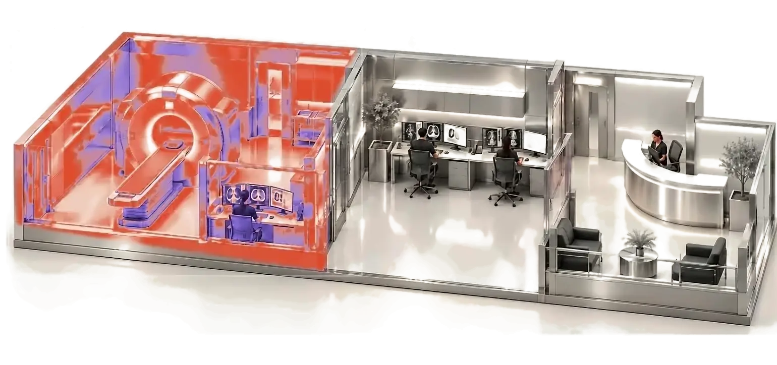
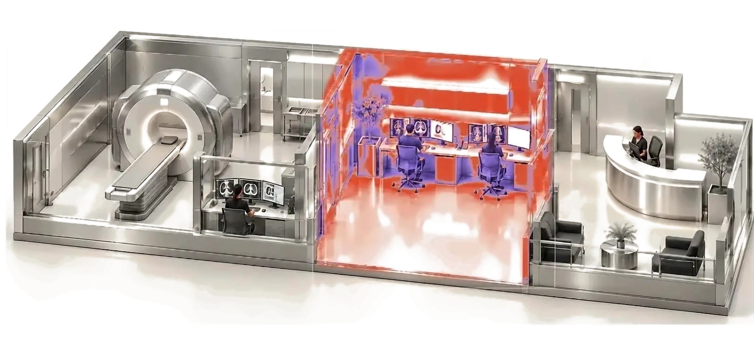
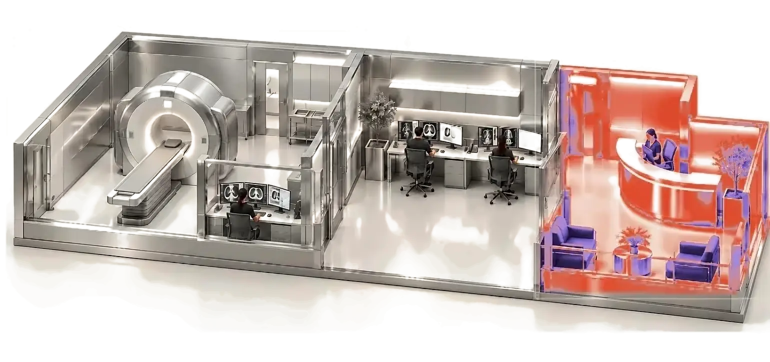
Your PACS and your RIS. Now connected.
Laudos.AI connects to the systems the clinic already uses, with no equipment swap and no pause in the exam schedule.
01 / 03Your voice. Your template. No typing.
You dictate as you speak. The report follows your template and uses your specialty's terminology.
02 / 03Report signed. Patient served.
Critical finding flagged for immediate communication and the report ready for release as soon as the physician signs. The queue moves and turnaround time drops.
03 / 03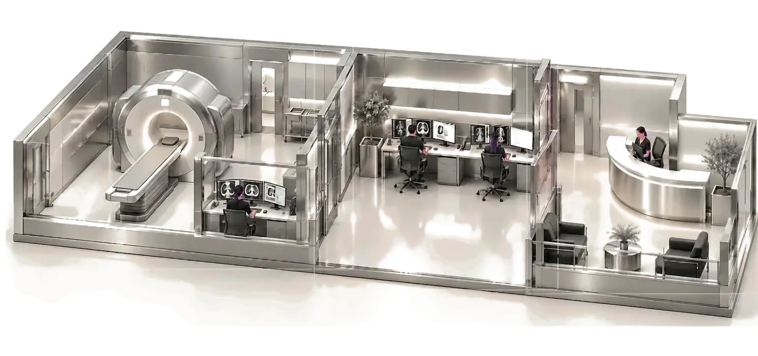
Laudos.AI RADAR · for institutions
The report is doneThe work isn't
RADAR flags reports that may need communication, peer review or follow-up. It also compares the report, billing and authorization to flag documentary claim-denial risk.
A critical finding needs more than a conclusion.
Find reports with findings from your critical-results list. Check whether communication is needed, and track contact and receipt as separate steps.
Intracranial hemorrhage
What the document saysAcute subdural hematoma.
In this example, no contact is recorded in the session yet. This does not prove that no communication occurred in another system.
A reason for review, not just a flag.
Compare technique, measurements, laterality, findings and conclusion. Locate possible inconsistencies and the passages that support referral for peer review.
Conflicting laterality
What the document saysKidney lesion, right side.
Kidney lesion, left side.
Check both statements before making a correction. Text analysis does not establish which side is correct.
The recommendation stays visible to the team.
Find recommendations for follow-up and further investigation in the report. Identify what needs review and follow-up, with the source passage available.
Follow-up recommended in the report
What the document saysSuggested: follow-up CT in 3 months.
The interval comes from this example document. The team assesses the recommendation in its clinical context.
Does the report support the billed procedure?
Compare the report, billing and authorization. Check differences in procedure, contrast and laterality before billing or during payer review.
Contrast: report versus billing
What the document saysExam performed without contrast (IV).
Chest CT with contrast (IV).
Documentary claim-denial risk. Check the administration record and reconcile the documents. No automatic approval or denial.
Who uses it
Professionals ending their shiftwith less friction
“I can report every exam within the shift itself. I no longer take any work home. And it also prevents rework.”
“I use Laudos.AI for my Doppler studies. The precision in structuring the findings is what stands out most.”
Pricing
Try it in your real routineDecide later
R$ 0
14 days or up to 30 reports. Your exams, your template, your routine. No card and no automatic charges.
Create a free accountR$ 299/month
Up to 1,000 reports per month. Structured dictation, classifications with their criteria and house templates.
Start with the free trialCustom
PACS/RIS integration, CRIT and RADAR with SLA, template governance and a live dashboard.
Book a demoResources
Guides to radiology reporting
Choose a guide for the step you want to improve: dictation, review or software selection. Each guide explains what to compare, how to test and the limitations of each feature.
Radiology reporting software
Report editors, speech recognition and AI report structuring handle different tasks. Compare the effort required to adapt your templates, review the text and submit reports in your official system. The guide covers cost, access and integration criteria for testing the software in your workflow.
Dictation for medical reports
Dictate terms, measurements and negations as you would during a shift. Then check what was transcribed, what went into the draft and what needed correcting. The guide distinguishes speech recognition from report structuring.
How to evaluate AI for medical reports
Compare the findings provided with every statement in the generated text. Record omissions, additions, laterality, measurements and structural changes. Refer to the review criteria and the limitations of the metrics before assessing time savings.
Questions and answers
Before youtry it
Questions about reporting, management and IT. If yours is not here, contact the team.
Talk to the teamNo. Laudos.AI supports reporting in radiology, nuclear medicine, cardiology and other specialties. You dictate the findings; the report follows your template. You review and sign.
Every result is an assistive draft. You check findings, measurements, laterality, classifications and conclusion. Interpretation, editing and signature remain the physician's, and every change is recorded.
14 days or up to 30 reports, whichever comes first. No card and no automatic charge. After that, the Pro plan costs R$ 299/month for up to 1,000 reports.
Integration depends on the systems and the scope. In the institutional demo, the team assesses your setup before defining the proposal.
Laudos.AI complies with CFM Resolution 2.454/2026 and the LGPD (Brazil's data protection law). The security documentation is ready for IT and DPO review.
More reports done. More life after.
You interpret. Get help with the writing. Try 14 days or 30 reports, no card required.